
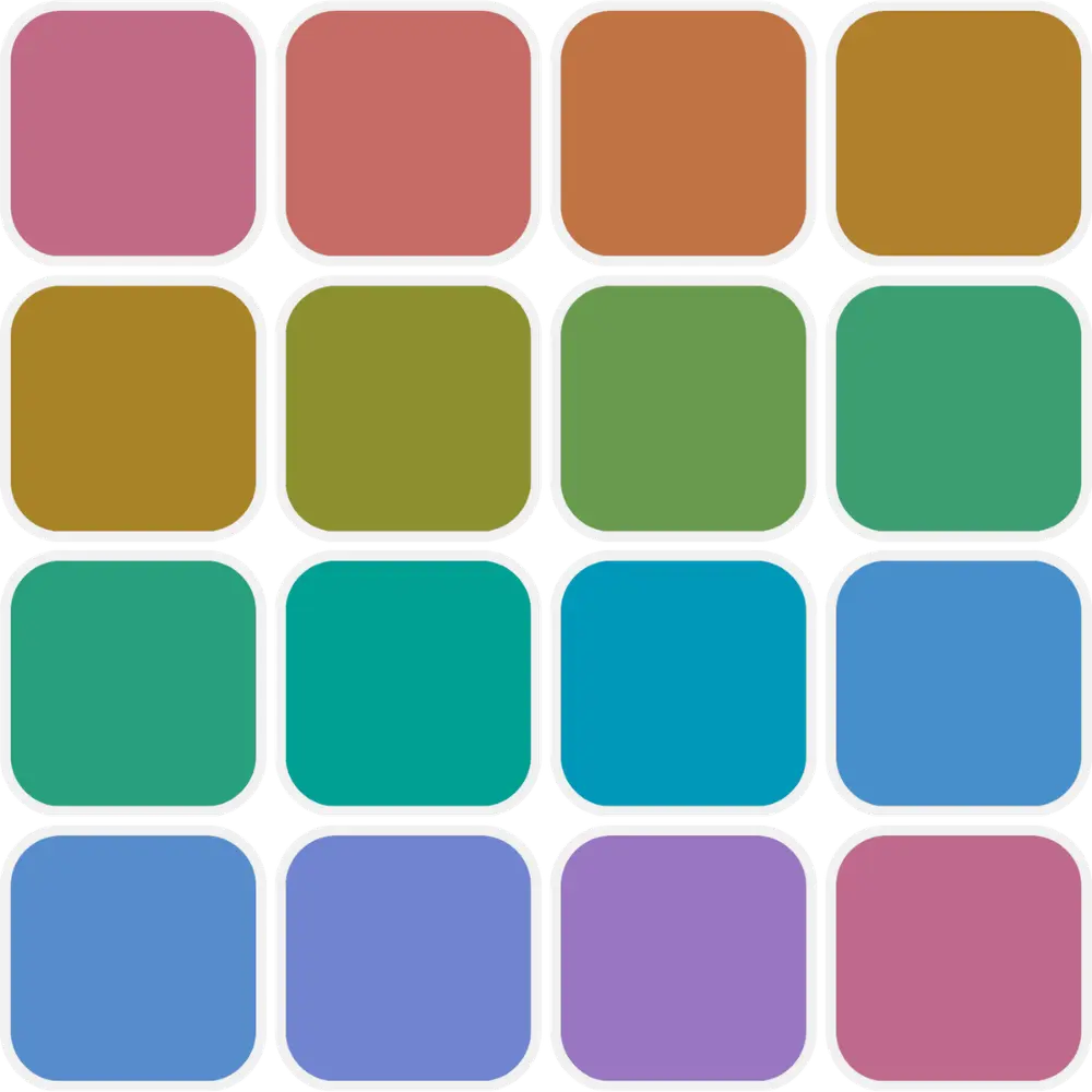
Yes. The BlindnessTest Farnsworth-Munsell 100 Hue Test is free in your browser with instant results and no sign-up required.
Farnsworth-Munsell 100 Hue Test
Take a free online Farnsworth-Munsell 100 Hue Test and measure how finely you can tell one hue from the next. You drag color caps into a smooth rainbow order between fixed anchors, and the tool scores each cap by how far it sits from its true neighbors.
Take the FM 100 Hue Test
Choose Quick (40 caps, all rows at once) or Full (85 caps, four trays). Drag the caps into smooth hue order between the fixed ends, then submit for your total error score and polar error diagram.
Most quick color vision screens ask a yes-or-no question: can you read the hidden number or not. The Farnsworth-Munsell 100 Hue Test asks something finer. It measures how precisely you can order colors that differ only slightly in hue, across the entire color circle rather than on the red-green axis alone. That makes it one of the most sensitive ways to reveal subtle discrimination difficulty, including mild cases that pass plate tests.
The test was created by Dean Farnsworth in 1943 and standardized in its familiar 85-cap form in 1957. The name says 100, but the physical kit uses 85 numbered caps of constant lightness and saturation that vary only in hue, arranged in four trays. In a clinic they are sorted under daylight-like Illuminant C, and the examiner scores how far each cap lands from its correct neighbors.
BlindnessTest keeps the arrangement task and the neighbor-based scoring on your everyday screen. Quick mode shows 40 caps in four rows at once for a fast screen. Full mode runs all 85 caps one tray at a time, closer to the clinical cap count. Both produce a total error score and a polar error diagram, but neither is a physical Munsell kit under controlled light, so treat the numbers as screening and education, not clinical grading.
If a red-green plate test looked normal but subtle shade sorting still feels hard, this is the tool that often shows it. Review test settings explained for Quick and Full modes and the timer before you start.
How Does the Farnsworth-Munsell 100 Hue Test Work?
You arrange movable color caps so the hue changes smoothly from one fixed anchor cap to the other. Because every cap shares the same lightness and saturation and differs only in hue, you cannot use brightness as a shortcut. The only way to order them correctly is to perceive the small hue steps between neighbors, which is the discrimination the test is probing.
When you submit, each cap is scored by how far its position sits from its true neighbors. The tool adds the distance to the cap on the left and the distance to the cap on the right, then subtracts two, so a cap sitting between its correct neighbors scores zero. Misplaced caps score higher, and a single swap raises the score of the caps around it. Adding every cap error gives your total error score, where lower is better.
Quick mode uses 40 caps, with 32 you can move and 8 fixed anchors, shown as four rows at the same time. Full mode uses all 85 caps, 77 movable and 8 anchors, presented one tray at a time like the clinical kit. Choose Quick or Full on the welcome screen, open Settings there to set an optional time limit, tile size, progress, and background, then drag the caps into order.
After you submit, a polar diagram plots your error at each position around the hue circle. A low, flat ring means even discrimination. Tall spikes clustered in one region show where neighboring hues were hardest to separate, which is how the test hints at a red-green or blue-yellow pattern.
How to Prepare for Hue Arrangement
Hue arrangement is sensitive to display quality and fatigue. These steps help the subtle cap-to-cap hue steps stay visible from the first row to the last. Choose Quick (40 caps) or Full (85 caps) on the welcome screen, and open Settings there to set an optional timer, tile size, and background before you start.
- Set screen brightness to a comfortable, steady level and keep it the same for the whole run.
- Turn off Night Shift, True Tone, blue-light filters, and color-correction or accessibility tint apps.
- Use even indoor or daylight-neutral lighting and reduce glare on the panel. Avoid strong colored lamps.
- Use a desktop or laptop with a color-capable display when possible. Phone screens can compress subtle hue steps.
- Remove tinted lenses for the run. Keep everyday prescription glasses on if you normally wear them.
- Complete all caps in one sitting without long breaks, since fatigue affects fine hue discrimination.
Test Settings Explained
Test length
Quick uses 40 caps shown as four rows at once and takes about three minutes, good for a fast screen. Full uses all 85 caps across four trays, one tray at a time, and takes roughly ten to twelve minutes for a run closer to the clinical cap count. Choose on the welcome screen before you start.
Time limit
The timer is off by default, so you can work at your own pace. You can also set 3, 5, 10, or 15 minutes if you want a timed run. Fine hue sorting is sensitive to fatigue, so a relaxed pace usually gives a cleaner result.
Tile size
Comfortable uses larger caps that are easier to compare and drag, which suits phones and touchscreens. Compact fits more caps on screen at once, which can help on smaller desktop windows. Open Settings on the welcome screen to switch before you begin.
Progress
Show displays a small tray counter (Tray X of 4) during the Full 85-cap test so you know how far you are. Hide removes it for a cleaner board. Quick mode shows all four rows at once, so there is nothing to count.
Background
Choose White or Black for the tray surround behind the caps. Black can make the subtle hue steps easier to judge for some people, while the caps themselves stay the same. Open Settings on the welcome screen to switch.
Fixed anchors
The caps at each end of every row or tray are locked in place. They mark the correct start and end hue so you always know which direction the sequence should flow. Only the caps between the anchors can be moved.
Drag to order
Pick up a cap and drop it into position so the hue changes smoothly from neighbor to neighbor. Keep rearranging until the whole row or tray looks like an even gradient. In Full mode, tap Next tray to continue, then Submit on the last tray. There is no penalty for adjusting as many times as you like.
Saved preferences
Your timer, tile size, progress, and background choices persist in local browser storage on the same device. This tool does not store your score between sessions, so note your total error score and polar pattern if you want to compare later.
Where Errors Cluster on the Hue Circle
The value of a hue arrangement test is not just the total score but where the mistakes fall. Each type of color vision deficiency makes a particular part of the color circle hard to order, so errors pile up in a predictable region. The polar diagram maps that pattern. Drag each slider below to see how the parts of the spectrum that cause trouble shift with protan, deutan, and tritan patterns.
Protan Pattern (Red Region)
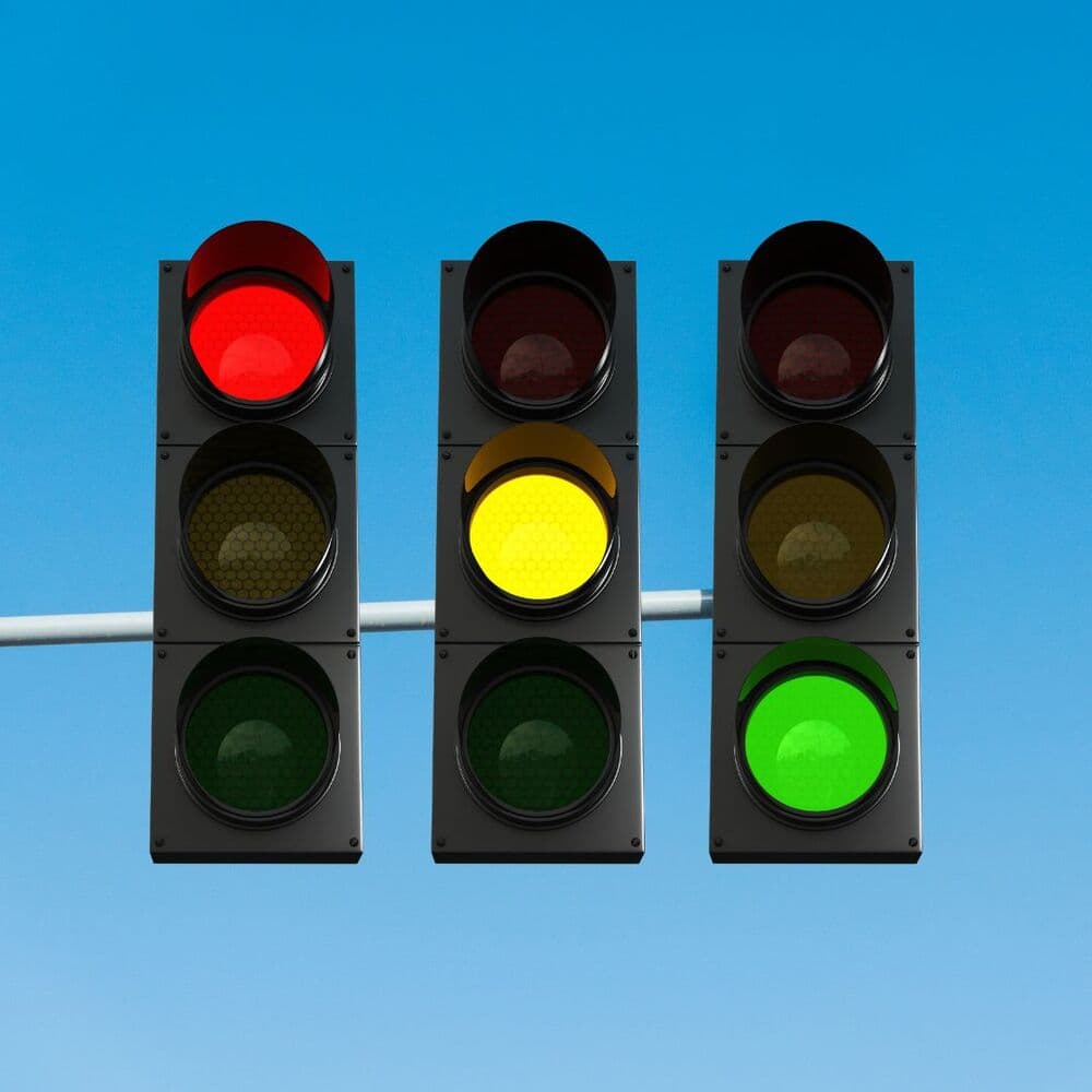
Protan patterns come from a shift or loss in the long-wavelength L cones. On the hue circle, errors tend to concentrate in the red to yellow region, where warm hues become harder to separate and reds can also look darker. Contrary to a common myth, the Farnsworth-Munsell test does detect red-green difficulty well; reading the error region is often more sensitive to mild loss than a plate pass or fail.
On the polar diagram a protan pattern shows a cluster of taller spikes in the red-orange segment rather than an even ring. The test does not label protanopia versus protanomaly. For a red-green versus blue-yellow split and cone-type hints, follow up with the Color Blind Test.
In everyday terms, a protan arrangement often means the warm colors that should step cleanly from red to orange to yellow bunch together instead, so sorting paint chips or reading resistor bands can feel like guesswork. The polar plot turns that lived experience into geometry: the caps you struggled to place become the tall spikes along the red-orange arc, and their height tracks how far each one landed from its true neighbors.
Deutan Pattern (Green Region)

Deutan patterns involve the medium-wavelength M cones and are the most common inherited red-green form. Errors tend to cluster in the yellow to green part of the hue circle, overlapping the protan region but shifted. Mild deuteranomaly is exactly the kind of subtle case a hue arrangement task can surface when quick plate screens read as normal.
Because the two red-green regions sit opposite each other on the circle, a deutan error cluster often appears as spikes on both the yellow-green and the far magenta-red segments. This bipolar pattern is a hallmark of red-green confusion on the polar plot, and it is why the diagram matters as much as the single total score.
Deutan observers frequently sort the blues and violets with ease and then stall in the greens, where several caps look like the same muted shade. Because it is the most common inherited pattern, it is also the one most often missed by a quick plate test that a mild deuteranomalous observer can still pass. That is exactly where a full hue arrangement earns its keep, since ordering precision reveals difficulty a pass-or-fail plate never records.
Tritan Pattern (Blue-Yellow Region)
Tritan patterns involve the short-wavelength S cones on the blue-yellow pathway. Congenital tritan deficiency is rare, but this axis is also sensitive to acquired changes from aging, medication, and eye disease, and the hue arrangement task is one of the better ways to reveal it. Errors cluster in the blue and yellow regions rather than the red-green regions.
On the polar diagram a tritan pattern places tall spikes in the blue-violet and yellow segments, roughly at right angles to a red-green cluster. If your errors concentrate there, or if blue-yellow tasks feel newly difficult, treat it as a reason to see an eye care professional rather than a diagnosis from this screen.
Unlike the red-green patterns, a tritan cluster is worth taking seriously as a possible acquired change rather than a lifelong trait. If your blue and yellow caps drift out of order and you have not noticed this before, note whether you take medication, have had recent changes in eye health, or are simply getting older, since all three can raise blue-yellow errors, then raise the pattern with an eye care professional.
The Hue Circle You Are Sorting
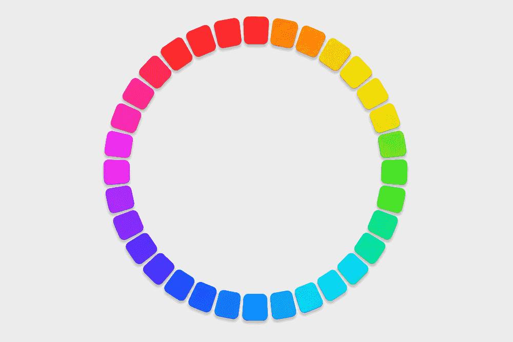
Every cap in the test shares the same lightness and saturation and differs from its neighbors only in hue, so together they form a smooth ring around the color circle. In the tool the ring is broken into rows or trays and the movable caps are shuffled, and your job is to rebuild the smooth transition between the fixed anchors.
The cap colors are placed at even steps around a perceptually uniform hue circle, so neighboring caps are genuinely close in hue. That even spacing is what makes the task a fair test of fine discrimination: if two adjacent hues look identical to you, that is where a mistake, and an error spike, will appear.
You never see the whole ring at once during the test. Quick mode breaks it into four rows and Full mode into four trays, and within each one the movable caps are shuffled before you begin. Rebuilding the smooth transition segment by segment is what turns a simple sorting task into a sensitive measure of how finely you can separate one hue from its neighbor.
Reading the Polar Error Diagram
Your result includes a polar diagram that plots the error at each cap position around the hue circle. Short spikes mean the caps near that hue were placed close to their correct order. Tall spikes mean larger displacements, so that part of the spectrum was harder for you to sort.
The shape tells the story. An even, low ring points to typical hue discrimination. A cluster of tall spikes in one area, often mirrored on the opposite side of the circle, points to a specific pattern: red-green clusters fall in the red and green regions, while blue-yellow clusters fall roughly at right angles to them. This is why the diagram is as informative as the total error score itself.
Reading the diagram is a two-step habit. First check whether the ring is broadly low and even or has clear peaks, then look at where any peaks sit. A single tall spike is often just one careless swap, while a run of tall spikes across one arc, mirrored on the far side, is the signature of a genuine axis pattern rather than a momentary slip of attention.
Why Screen Color Shifts Your Cap Order
A clinical Farnsworth-Munsell kit uses physical Munsell caps viewed under daylight-like Illuminant C. Your phone, tablet, or monitor renders the caps as sRGB colors under whatever light is in your room, so the hue steps you see are not identical to the printed standard. Cap colors here are generated on a perceptually even hue circle, which keeps the task fair, but it is an approximation of the physical papers, not a match.
Set brightness to a comfortable level, turn off Night Shift, True Tone, and blue-light filters, remove tinted lenses, and reduce glare before you start. A larger, color-capable screen usually shows subtle hue steps better than a small phone. Because conditions vary, use the same device and settings if you want to compare runs over time, and treat a single score as a screening signal rather than a fixed measurement.
Frequently Asked Questions
Limitations of the Online Farnsworth-Munsell 100 Hue Test
Screening, not diagnosis
This online arrangement task is for screening and education. It does not diagnose color blindness, separate protanopia from protanomaly, or replace a comprehensive exam by an eye care professional.
Plate tests vs hue arrangement
Ishihara and similar plate tests screen common red-green problems quickly with a pass-or-fail style result. This test quantifies fine hue discrimination across the whole circle and maps where errors cluster, including red-green regions. Use both when you want fast screening plus hue detail.
Online vs clinical kit
A professional kit uses 85 physical Munsell caps under controlled Illuminant C with supervised scoring. Full mode follows the 85-cap tray layout on an uncalibrated display, and Quick is a shorter 40-cap screen. Neither matches instrument-grade cap standards or published norms exactly.
Occupational certification
Aviation, maritime, rail, and other regulated fields often require in-person color vision testing with approved protocols. The Farnsworth manual itself states the test was not designed as a simple pass-or-fail licensing screen. This tool does not certify you for licensing or hiring.
Academic References and Clinical Context
BlindnessTest scores your arrangement with the standard Farnsworth neighbor-error method: for each cap, add the distance to its left and right neighbors and subtract two, then sum across all caps for a total error score where lower is better. Errors are also grouped into red-green and blue-yellow cap regions to hint at which axis is affected, and plotted on a polar diagram.
The references below connect the hue arrangement paradigm, its scoring mathematics, and its normative interpretation to this screener. Results still reflect performance on your uncalibrated display and do not replace a physical Munsell kit under controlled light or a comprehensive eye examination.
- Farnsworth (1943)
The Farnsworth-Munsell 100-Hue and Dichotomous Tests for Color Vision. Journal of the Optical Society of America, 33(10), 568-578. The original description of the hue arrangement task and its scoring that this tool adapts.
- Indian J. Ophthalmology review
A review of Farnsworth-Munsell 100 Hue scoring and interpretation, including the per-cap neighbor-error formula and total error score used here. PMC4131328.
- Birch et al. (1992)
Updated normative data for the FM100 across ages 5 to 79. Establishes that total error score rises with age, so score bands must consider age and first-versus-repeat testing.
- Rodriguez-Carmona et al. (2016)
Using the FM100 to distinguish protan and deutan observers from error patterns. Supports reading the polar error region, not just the total score, to hint at axis.
- Esposito & Houser (2018)
An adjusted error-score calculation for the FM100 under non-standard illuminants. LEUKOS. Explains why lighting and display, which are never true Illuminant C online, affect cap order and scoring.
- FM100 vocational scoring (1995)
Scoring the FM100 for vocational guidance. PMID: 8539021. Shows a total error score under 100 passes most normals, and that individual cap errors in red-green regions can flag mild difficulty even when the total looks average.
- National Eye Institute (NEI)
Explains inherited and acquired color vision deficiency for patients, including the acquired blue-yellow changes that hue arrangement tasks are well placed to surface.
- American Academy of Ophthalmology (AAO)
Ophthalmology-reviewed patient guidance on color blindness symptoms and care, supporting our position that an online arrangement score is screening, not diagnosis.
- American Optometric Association (AOA)
Covers symptoms, professional testing options, and follow-up with an optometrist when screening suggests a hue discrimination difficulty.
How sharp is your color perception?
Arrange the color caps in hue order with the Farnsworth-Munsell 100 Hue Test.