
Yes. The BlindnessTest Anomaloscope Test is free in your browser with instant results and no sign-up required.
Anomaloscope Test
Take a free online color matching test inspired by the clinical anomaloscope. Move a slider until both halves of a split disc look the same, then choose Match or Match Not Possible for each of seven pairs. Choose a red-green or blue-yellow (tritan) test with the start buttons on the welcome screen.
Take the Anomaloscope Test
Follow the on-screen prompts to complete the screening. Your result appears as soon as you finish.
Anomaloscope Test
Match the colors on both sides of the disc. Choose Match Not Possible if nothing looks right.
Traditional clinic matching tests use special lights and trained examiners to record where along a color axis a person accepts a match. Those devices work well in controlled rooms but are not available at home. On a phone, tablet, or monitor, fixed screen colors behave differently from clinical light sources because of display backlights, viewing angles, and subpixel layouts.
BlindnessTest adapts the matching idea to your screen with seven fixed color pairs and one brightness slider. Instead of a hidden numeral on a plate, you decide whether each pair looks matchable along one axis. The tool counts how many pairs you accept and gives a screening label on the red-green or blue-yellow test type you choose.
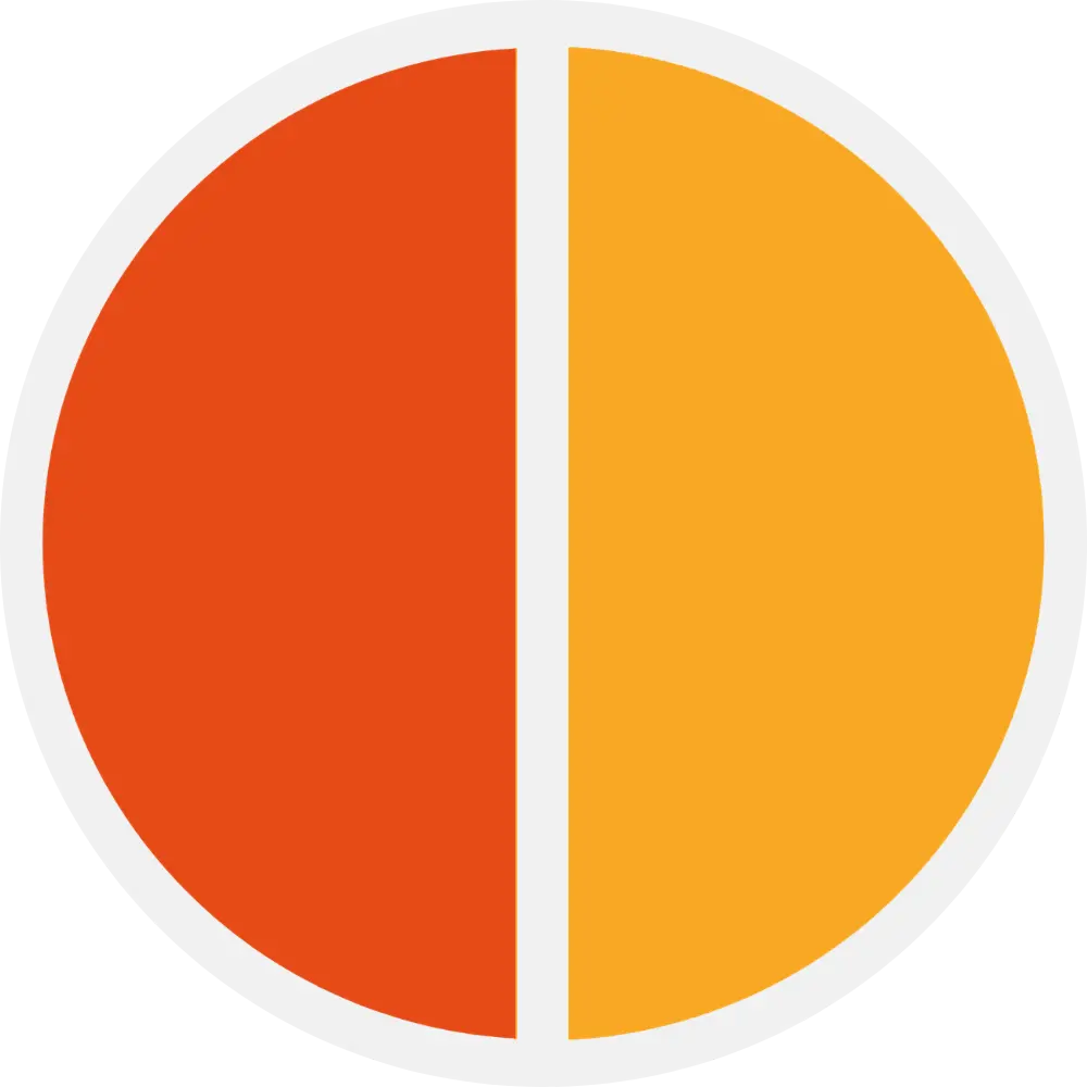
Each run shows a circle split into two halves. Move the slider until both sides look identical, then tap Match. If no setting looks right, tap Match Not Possible. The slider keeps its position when the next pair loads. Pair one is always the control reference. Pairs two through seven use shuffled references from a fixed palette.
If you have only used Ishihara plates or the Color Blind Test before, this adds a different follow-up. Ishihara screens with hidden figures. The Color Blind Test scores red, green, and blue channels separately. Choose Start Red-Green Test or Start Tritan (Blue) Test on the welcome screen, review test settings explained for display options, or read what your results mean after you finish.
How Does the Anomaloscope Test Work?
Color matching tests look at how broadly you accept different mixtures along one color axis. People with typical color vision usually match only the control pair that truly belongs on the diagonal. People with red-green or blue-yellow difficulty often accept extra pairs that do not truly match because their cone signals overlap along that axis.
Each run shows seven pairs. Pair one is the control on the diagonal. Pairs two through seven use shuffled reference colors from a fixed palette. You adjust one slider on the diagonal until both halves look alike, or you report that no match is possible. The red-green test type uses a yellow-style ramp. The blue-yellow (tritan) test type uses a cyan-style ramp.
When all seven pairs are done, the summary compares your answers against the rules in the scoring table under What Your Results Mean. If you accept only the control pair with a precise slider setting, screening reads as normal. If you accept more pairs, the label moves through mild, medium, or severe bands on the axis you selected. An imprecise control match or zero accepted matches may suggest a setup issue or inconsistent answers rather than a final clinical label.
How to Prepare for Color Matching
Color matching pairs respond to how your screen renders hue. These steps help your pairs stay consistent from run to run. Choose Start Red-Green Test or Start Tritan (Blue) Test on the welcome screen, and open Settings there for the pairs counter and background before you start.
- Set screen brightness to 100% before you start.
- Turn off night mode, blue-light filters, True Tone, and color-correction or accessibility tint apps.
- Remove tinted or colored lenses. Keep everyday prescription glasses on if you normally wear them.
- Use your best-quality display when possible. Results can differ between phones, tablets, and monitors.
- Sit in even indoor light, finish in one sitting, and take your time on each match decision.
- View the screen straight-on at your usual distance. Avoid glare and keep the display flat.
What Your Anomaloscope Results Mean
Your result counts how many of seven pairs you accepted as matches on the red-green or blue-yellow variant you chose when you started the run. Labels describe screening severity on that axis only. They do not separate protan from deutan and are not clinical instrument scores.
The first pair is the control. If you match only that pair and control precision Δ is below 10, the modal labels the run Normal. If you match the control but Δ is 10 or higher, the result notes imprecise matching. Retest in ideal lighting if that surprises you.
When you accept two or more pairs, severity follows the match count in the table above: mild at two, medium at three to four, and severe at five to seven. Medium and severe names include the red-green or blue-yellow axis from the variant you started, not protanopia versus deuteranopia.
The result modal shows a match profile strip (numbered circles for all seven pairs) plus control precision in the summary when you matched the control. This online tool uses fixed on-screen colors and one brightness slider, not spectral lights and two clinical knobs.
For cone-type hints and channel percentages, continue with the Color Blind Test. For occupational or medical certification, confirm with an in-clinic exam under your regulator's required protocol.
Match-count screening on seven pairs (one control pair plus six reference pairs)
| Pairs you accepted | Screening label | What it means on this site |
|---|---|---|
| 0 | Invalid | The control pair should be matchable. Retest with stable lighting and check display settings. |
| 1 with precise control | Normal | Only the control pair accepted with a close slider setting. Typical screening on the test type you chose. |
| 1 with imprecise control | Imprecise match | Only the control accepted, but the slider was far from the reference. Retest recommended. |
| 2 | Mild | Red-green or blue-yellow axis label from the variant you ran. Not protan versus deutan typing. |
| 3 to 4 | Medium | Broader acceptance of extra reference pairs along the chosen axis. |
| 5 to 7 | Severe | Many extra pairs accepted on this RGB matching task. |
The Split Disc You Are Matching
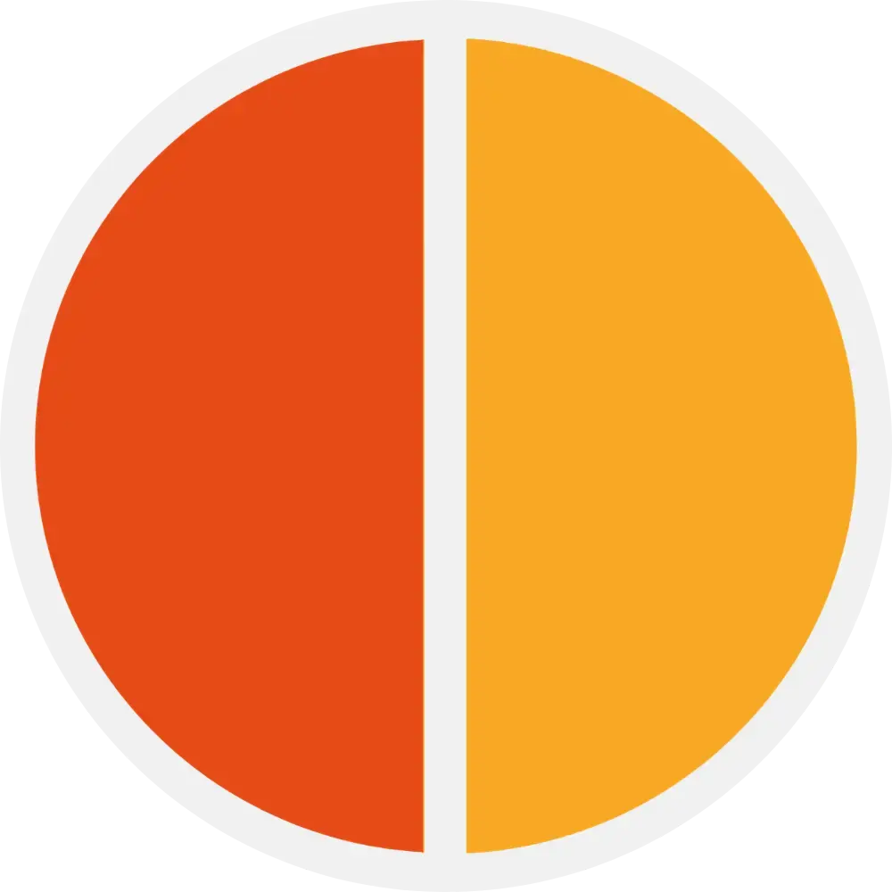
The clinical Rayleigh match, first described by Lord Rayleigh in 1881, asks you to mix red and green light until it matches a fixed yellow. The red-green mode borrows that idea: one half is a mixture you control with the slider, and the other half is a fixed reference. Because a red-green deficient eye weighs red and green differently, the mixture that looks like a perfect match to you can reveal how your cones combine those wavelengths. The blue-yellow (tritan) mode swaps in a simplified cyan-axis match rather than a red-green mixture.
On a clinical instrument the two halves are narrow-band lights and the examiner records the exact midpoint you choose. Here the halves are sRGB colors and you simply accept or reject each pair, so the disc keeps the matching idea while staying honest about being a screen, not a spectral device.
Test Settings Explained
Test variant
Red-green uses a yellow-style matching axis for common red-green screening. Blue-yellow (tritan) uses a simplified cyan axis for blue-yellow screening. Choose Start Red-Green Test or Start Tritan (Blue) Test on the welcome screen. The choice is locked once the run begins.
Seven pairs
Every run uses one control pair plus six shuffled references. You always complete all seven before the result modal opens. There is no timed pressure. Take your time on each match decision.
Slider persistence
The slider keeps its value between pairs while the reference half changes. You can still move it freely on each pair. Each run starts with the slider visibly off the control match so both halves look different at first.
Pairs counter
Show displays the current pair number during the run. Hide removes the counter for a cleaner view. The counter shows progress through the seven-pair sequence, not a score.
Background
Choose White or Black for the area behind the split disc. Black can make the two halves easier to compare in a dim room. Open Settings on the welcome screen to switch before you start.
Match buttons
Tap Match when both halves look the same. Tap Match Not Possible when no slider position looks acceptable. Only accept a pair when the match looks genuine.
Saved preferences
Your last variant, pairs counter, and background choices persist in local browser storage on the same device. Open Settings on the welcome screen to adjust the pairs counter and background before you start.
Red-Green Color Vision and Matching

Most inherited color vision deficiency affects red-green discrimination. Matching tests probe that axis by asking how many extra reference pairs still look acceptable. Drag the slider to compare how a familiar scene can look with normal color vision versus deuteranopia.
Red-green deficiency is the most common form of color blindness, inherited on the X chromosome and primarily affecting males. When you choose the red-green test type, this tool reports how many extra pairs you accept along that axis. It does not label protan versus deutan. Only an in-person exam with calibrated equipment can confirm subtype and severity.
The reason matching reveals red-green deficiency so cleanly is that it never asks you to name a color, only to judge when two fields look the same. Naming can be coached or guessed, but a match either holds or it does not. When your red and green cones carry overlapping signals, mixtures that look clearly different to a typical observer can settle into an apparent match, and the extra pairs you accept are exactly that effect made countable.
Protan Patterns in Daily Vision
Protan patterns involve reduced sensitivity on the red-cone axis. Clinical matching equipment can sometimes separate protan from deutan under supervision using detailed records. This online tool uses the same fixed references for everyone and does not emit those clinical subtype labels.
The word protan points to the long-wavelength L cones specifically. In a clinical Rayleigh match, protan observers give themselves away not only by accepting a wide range of red-green mixtures but by needing the yellow reference dimmed, because red light itself looks darker to them. That brightness clue is one of the details a screen version cannot fully reproduce.
On this tool you will not see that luminance signature directly, since one slider and fixed pairs stand in for the clinical knobs. What you can see is breadth: a protan pattern tends to accept several mixtures that a typical observer would reject outright.
If Ishihara or this matching screen suggests difficulty and you want protan or deutan hints, continue with the Color Blind Test or discuss formal testing with an eye care professional.
Blue-Yellow Deficiencies (Tritan)
Tritan deficiencies affect the short-wavelength (blue) S cones. This form is much rarer than red-green deficiency. Some people with tritan involvement still pass red-green matching in clinic, so blue-yellow screening needs its own test type.
Choose Start Tritan (Blue) Test on the welcome screen to run the simplified cyan axis on this site. Results describe how many pairs you accept on that axis. They are screening bands only, not formal clinic scores from a calibrated instrument.
Blue-yellow matching draws on a different pair of cones than the red-green axis, so a normal red-green result tells you nothing about it. That is why the variant is a deliberate choice on the welcome screen rather than something the tool decides for you.
Because congenital tritan deficiency is so rare, a raised blue-yellow result more often points to an acquired cause: the natural yellowing of the lens with age, some medications, or conditions affecting the retina and optic nerve. None of that can be diagnosed from a browser, but a consistent blue-yellow pattern here is a sensible reason to book a full eye examination rather than to worry in the abstract.
How Match Count Relates to Severity

On clinical matching equipment, mild difficulty often means accepting a slightly wider range of mixtures. More severe difficulty can mean accepting matches across most of the axis. This online tool approximates that idea by counting how many pairs you accept as a match in total, including the control pair.
Accepting only the control pair reads as normal. Two accepted pairs suggest mild screening on this scale, three to four suggest medium, and five to seven suggest severe on this screen-based task. These bands are estimates for awareness only. Repeat with stable lighting if results surprise you.
The logic behind the bands mirrors how a clinician reads a Rayleigh match: counting accepted pairs is a simple stand-in for measuring how wide a range of mixtures you are willing to call a match. The section below explains why that acceptance width, rather than any single slider position, is the number that matters.
Treat the boundaries as soft. One extra accepted pair can be the difference between a normal and a mild label, and glare, a warm screen filter, or a rushed decision can nudge you across a line. If a result surprises you, the most useful next step is not to retake immediately but to fix the viewing conditions first, then run it once more.
Why Acceptance Width Matters
The single most telling number in a matching test is not the exact point you pick but how wide a range of mixtures you are willing to call a match. Someone with typical color vision accepts only settings very close to the true match, a narrow band around the center of the axis. This tool's control pair checks that narrow acceptance.
Red-green deficiency widens that band. When several very different mixtures all look acceptable, the extra pairs you accept in this screening stand in for the wide matching range a clinician would measure on an instrument. That is why the label depends on how many pairs you accept in total, not on any single slider position.
This is not a new idea. The first anomaloscope was built by Willibald Nagel in 1907 around Lord Rayleigh's red-green match, and clinical instruments since then mix light near 545 and 670 nanometres against a 589 nanometre yellow. The classic reading is straightforward: a narrow acceptance range points to typical color vision, a wider but still limited range to anomalous trichromacy, and a match across the entire range to dichromacy, where one cone type is effectively absent. This screen approximates that same spectrum of outcomes with counted pairs instead of narrow-band lights.
Frequently Asked Questions
Limitations of the Anomaloscope Test
Screening, not diagnosis
Browser-based matching counts cannot replace calibrated clinical instruments, a full eye health exam, or occupational certification protocols.
Match count only
This tool reports how many pairs you accept on fixed screen colors. It does not output clinical instrument scores from a supervised matching exam.
No protan versus deutan typing
Results describe red-green or blue-yellow axis breadth from your test type choice. They do not classify protanopia versus deuteranopia. Use the Color Blind Test for channel scores and type hints.
Licensing and employment
Aviation, maritime, and medical licensing require supervised in-clinic testing. Use this result as a private screening step only.
Academic References and Clinical Context
Clinical matching tests use controlled lights and trained examiners to record where along a mixture axis a person accepts a match and how wide that acceptance is. BlindnessTest adapts the matching paradigm to fixed on-screen color pairs and a single brightness slider on your display.
The references below connect peer-reviewed vision science and patient guidance to this screener, including online display limitations and responsible result language. Results still reflect performance on your uncalibrated screen and do not replace calibrated in-clinic exams or occupational certification protocols.
- National Eye Institute (NEI)
Overview of inherited and acquired color vision deficiency. Explains types, daily impact, and when to seek professional evaluation. Helps frame how BlindnessTest describes red-green and blue-yellow screening labels.
- PubMed: Anomaloscope
Search peer-reviewed literature on clinical color matching tests and congenital red-green classification in research and clinical settings.
- American Academy of Ophthalmology (AAO)
Patient-focused explanation of color blindness types, symptoms, and clinical care. Aligns with our disclaimers: online results screen display performance and do not replace calibrated in-clinic exams.
- American Optometric Association (AOA)
Covers symptoms, professional testing options, and follow-up with an optometrist. In-clinic matching tests are one option providers may choose when detailed axis assessment is needed.
- NHS: Color Vision Deficiency
Guidance on red-green, blue-yellow, and complete color vision deficiency. Covers daily-life and vocational impact. Informs practical result summaries, not occupational certification or a formal diagnosis.
Want a matching-style screen?
Try the Anomaloscope Test for hands-on color matching on red-green or blue-yellow axes. Results count accepted pairs for screening, not protan versus deutan typing.